
 (6 votes, moyenne: 4.33 sur 5)


The use of mini screws as temporary anchorage devices has revolutionized orthodontics. The following case illustrates a type of orthodontic correction which was impossible to consider without surgery just a few years ago.
Case history :
- This 25 year old woman had started an orthodontic treatment 2 years earlier before being to Dr. John Graham orthodontist, practicing in Litchfield Park, Arizona.
- One of the patient’s chief complaint was that she didn’t like her gummy smile. The first series of photographs shows the severity of that gummy smile; more than 1 cm (10 mm) of gingiva is visible upon smiling.
- The initial treatment plan included a “maxillary impaction” which is an orthognathic surgery procedure aimed at “moving up” the upper jaw to reduce the gummy smile.
- Having lost her insurance coverage, the patient could no longer afford the surgical fee so the initial treatment plan had o be modified. Dr Graham offered to use temporary anchorage devices (TADs) to intrude the maxillary dentition and correct the gummy smile.
- The photos (A, B, C) below show the patient as she was transferred to Dr. Graham. (D) At the end of treatment, after the use of TAD mini-screws and reduction of the gummy smile.
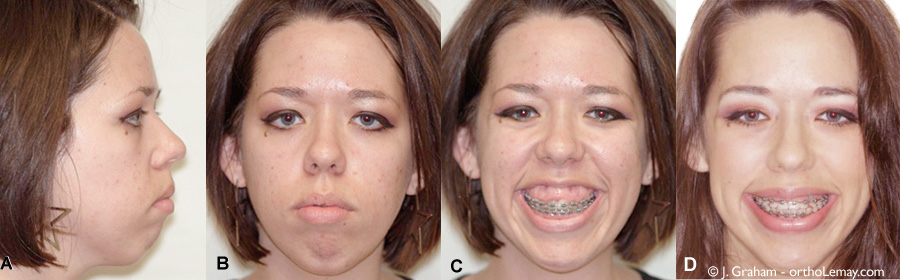
10 mm gummy smile decreased by orthodontic intrusion with mini-screw anchorage (TADs). (A, B, C) Occlusion when the case was transfered to Dr. Graham.. (D) At the end of treatment, after the use of mini-screws to reduce the gummy smile.
-
Although the occlusion and tooth alignment were acceptable at this stage of treatment (transfer), the smile still showed a lot of gingiva because this correction was originally planned to been done surgically. Treatment goals were still aimed at reducing the excessive gummy smile.

Acceptable occlusion prior to orthodontic intrusion using mini-screws.
Orthodontic mechanics used for the intrusion of the maxillary dentition

4 mini screws provided the necessary anchorage for the intrusion mechanics aimed at intruding the upper dentition and reducing the “gumminess” of the smile.
Cone Beam Computer Tomography (CBCT) imaging

Images from a Cone Beam Computer Tomography (CBCT) showing the position of the mini-screws (arrows). (A) Views of right, center and left sides. (B) Occlusal view showing the mini-screws positioned between the roots.
Cone Beam Computer Tomography (CBCT). Initial and final stages.
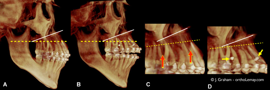
Vertical changes in the position of the teeth visible as shown in a 3D scan (Cone Beam Computer Tomography (CBCT)) before and after the intrusion.
To learn more about Cone Beam Computer Tomography imaging (CBCT)
Final results

(A) Final profile. (B) Front view and (C) smiling after orthodontics. The gummy smile is gone, no surgery!

Final occlusion

(A) Teeth during treatment, (B) at the end of treatment and (C) after the gingiva has been laser reshaped to give it a better contour. A dental facette was made by the general dentist on the upper right central incisor.
To learn more about soft tissue laser and how it can be used to modify gingival contour and improve esthetics.

(A) Severe gummy smile showing 1 cm of gingiva before treatment. (B) At the end of treatment. (C) After the installation of a dental facette on a central incisor. Note that a surgical procedure was done to modify the chin (genioplasty).
To learn more about genioplasties
Thanks to Dr. John Graham for sharing this case.